Automated Tissue Image Analysis in Cancer Diagnostics: Transforming Precision Medicine
Automated tissue image analysis or histopathology image analysis (HIMA) is the use of computer-controlled automatic test equipment to evaluate tissue samples, using computations to derive quantitative measurements from an image to avoid subjective errors
3/26/20254 min read
Introduction
The integration of artificial intelligence (AI) and machine learning (ML) in cancer diagnostics has revolutionized pathology by improving accuracy, efficiency, and reproducibility. One of the most promising advancements in this field is automated tissue image analysis, which enhances the ability to assess cancerous tissues with unparalleled precision. This technology enables the quantification of various biological markers, providing insights into tumor behavior, prognosis, and potential treatment pathways.
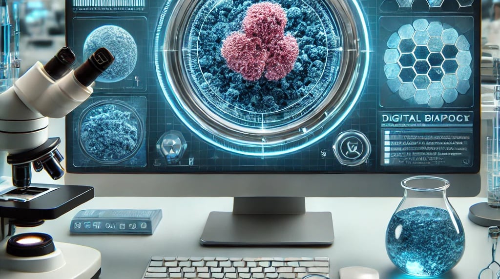
In clinical applications, automated tissue image analysis is crucial for evaluating the aggregate activity of cancer cells in biopsy samples. In breast cancer, for instance, this technology facilitates the detection of protein biomarkers that indicate tumor aggressiveness. By automating such assessments, pathologists and oncologists can make more informed decisions about a patient’s treatment strategy.
The Role of Automated Tissue Image Analysis in Cancer Diagnostics
Traditionally, pathologists examine biopsy samples under a microscope to assess cellular morphology, count tumor cells, and determine biomarker expression. While effective, this manual process is time-consuming and susceptible to variability. Automated tissue image analysis eliminates these limitations by using advanced algorithms to rapidly and consistently analyze histopathological images.
How It Works
Automated tissue image analysis employs a combination of digital pathology, computer vision, and machine learning models to:
Segment and classify different cell types within a biopsy sample
Quantify biomarker expression
Identify morphological changes associated with cancer progression
Generate comprehensive reports for pathologists and oncologists
By leveraging deep learning-based neural networks, these systems can recognize subtle patterns in tissue architecture and molecular expression that may not be easily discernible by the human eye.
Phases of Automated Tissue Image Analysis
The workflow of automated tissue image analysis typically consists of the following phases:
1. Image Acquisition and Digitization
The process begins with the digitization of tissue slides using high-resolution whole-slide imaging (WSI) scanners. This transformation allows pathology labs to store, share, and analyze images computationally, providing a foundation for AI-powered analysis.
2. Preprocessing and Segmentation
Before analysis, the system applies image preprocessing techniques such as noise reduction, contrast enhancement, and color normalization. Following this, image segmentation algorithms classify different regions of interest (ROIs) such as tumor areas, stromal tissue, and necrotic zones.
3. Feature Extraction and Classification
Using deep learning and statistical methods, the software extracts critical features such as:
Cell morphology (size, shape, nuclear atypia)
Biomarker expression levels
Tissue architecture patterns
These extracted features are then classified using machine learning models to differentiate between benign and malignant cells and identify cancer subtypes.
4. Biomarker Quantification
Automated analysis enables precise quantification of key protein markers, which is essential for predicting tumor behavior and treatment response. For breast cancer, for example, the system can assess:
Estrogen Receptor (ER) and Progesterone Receptor (PR) levels
HER2 (Human Epidermal Growth Factor Receptor 2) overexpression
Ki-67 proliferation index, an indicator of tumor growth rate
By accurately measuring these markers, oncologists can tailor therapies such as hormone treatments or targeted therapies (e.g., trastuzumab for HER2-positive breast cancer).
5. Report Generation and Clinical Interpretation
Finally, the system generates a structured diagnostic report, highlighting key findings, quantification scores, and potential clinical implications. Pathologists can review and validate these reports before finalizing their diagnosis.
Applications in Breast Cancer Diagnosis and Prognosis
Breast cancer is a heterogeneous disease with distinct molecular subtypes, necessitating precise diagnostic techniques. Automated tissue image analysis plays a pivotal role in:
1. Identifying Tumor Aggressiveness
AI-powered analysis can quantify HER2 overexpression, a critical factor in determining the need for HER2-targeted therapies.
High Ki-67 levels indicate a rapidly proliferating tumor, guiding decisions on chemotherapy intensity.
2. Predicting Treatment Response
ER/PR status determination via automated analysis helps identify candidates for endocrine therapy.
Digital quantification of tumor-infiltrating lymphocytes (TILs) provides insight into immunotherapy response likelihood.
3. Enhancing Reproducibility in Pathology
Automated quantification reduces observer variability among pathologists, ensuring consistent and reproducible results.
Available Software for Automated Tissue Image Analysis
Several AI-driven platforms have been developed for automated tissue analysis, including:
Paige AI – AI-powered pathology software for cancer diagnosis.
PathAI – Enhances accuracy in histopathological assessment.
Philips IntelliSite Pathology – Digital pathology system integrating AI analysis.
Visiopharm – Image analysis tools for cancer biomarker quantification.
Definiens Tissue Studio – Advanced tissue phenotyping software.
These platforms are integrated into clinical workflows, providing pathologists with enhanced diagnostic capabilities.
Implications and Limitations
Implications
Increased Efficiency: Automated systems analyze thousands of images in a fraction of the time required for manual examination.
Precision Medicine: Objective biomarker quantification improves treatment selection and patient outcomes.
Scalability: Digital pathology enables global collaboration, facilitating remote second opinions and telepathology.
Limitations
Data Quality Dependence: AI performance relies on the quality of histological staining and image acquisition.
Computational Challenges: High-resolution imaging requires substantial computing power and data storage.
Regulatory and Validation Hurdles: AI-based diagnostics require extensive clinical validation and regulatory approvals before widespread adoption.
The Future of Automated Tissue Image Analysis
The future of cancer diagnostics lies in the continuous evolution of AI-powered pathology. Key developments on the horizon include:
Integration with Multi-Omics Data: Combining histopathological analysis with genomics, proteomics, and metabolomics will offer deeper insights into cancer biology.
Real-Time AI-Assisted Diagnostics: Pathologists will receive instant AI-generated insights during live tissue examinations.
Augmented Reality (AR) in Pathology: AR-assisted microscopes will overlay AI-derived annotations onto tissue samples in real-time.
Improved AI Generalization: Future AI models will be trained on more diverse datasets to enhance their ability to handle varied tissue morphologies.
Conclusion
Automated tissue image analysis represents a paradigm shift in cancer diagnostics, offering precision, speed, and consistency in histopathology. For breast cancer patients, this technology provides quantifiable insights into tumor aggressiveness, aiding in personalized treatment planning. While challenges remain in regulatory approval and computational infrastructure, the trajectory of AI in pathology is set to redefine cancer care. With continued advancements, automated tissue analysis will become an indispensable tool in the fight against cancer, bridging the gap between digital pathology and precision oncology.
References
Williams, C., et al. (2021). "Advancements in AI-driven Histopathology: The Role of Automated Tissue Image Analysis." Journal of Digital Pathology.
Bush, K., et al. (2016). "AI and Biomarker Quantification in Breast Cancer: A Systematic Review." Nature Medicine.
Lauschke, V. M., et al. (2020). "Machine Learning in Pathology: The Road to AI-Augmented Diagnosis." Clinical Cancer Research.
Philips Healthcare. (2022). "Digital Pathology and AI: Transforming Cancer Diagnosis." Philips Whitepaper.
WHAT WE DO
MedTechSolns aims to provide credible, evidence-based, and technology-focused insight into healthcare systems, medical devices, diagnostics, and digital health innovations.
Our editorial approach bridges the gap between:
Medical science and technology
Clinical practice and health systems
Policy development and procurement decisions
Innovation and real-world implementation
The platform is designed to support healthcare professionals, policymakers, engineers, investors, and health system leaders in making informed decisions.
Resources
Connect
info@medtechsolns.com
+1234567890
© 2025. All rights reserved.